

Facility

Advanced Microscopy Unit

Provides technical and scientific support for high-resolution optical and fluorescence microscopy. Enables qualitative and quantitative analyses from tissues to subcellular structures, ensuring high-quality imaging with standardized protocols.

records

Operational records

Explore the resources and capabilities available for this service.

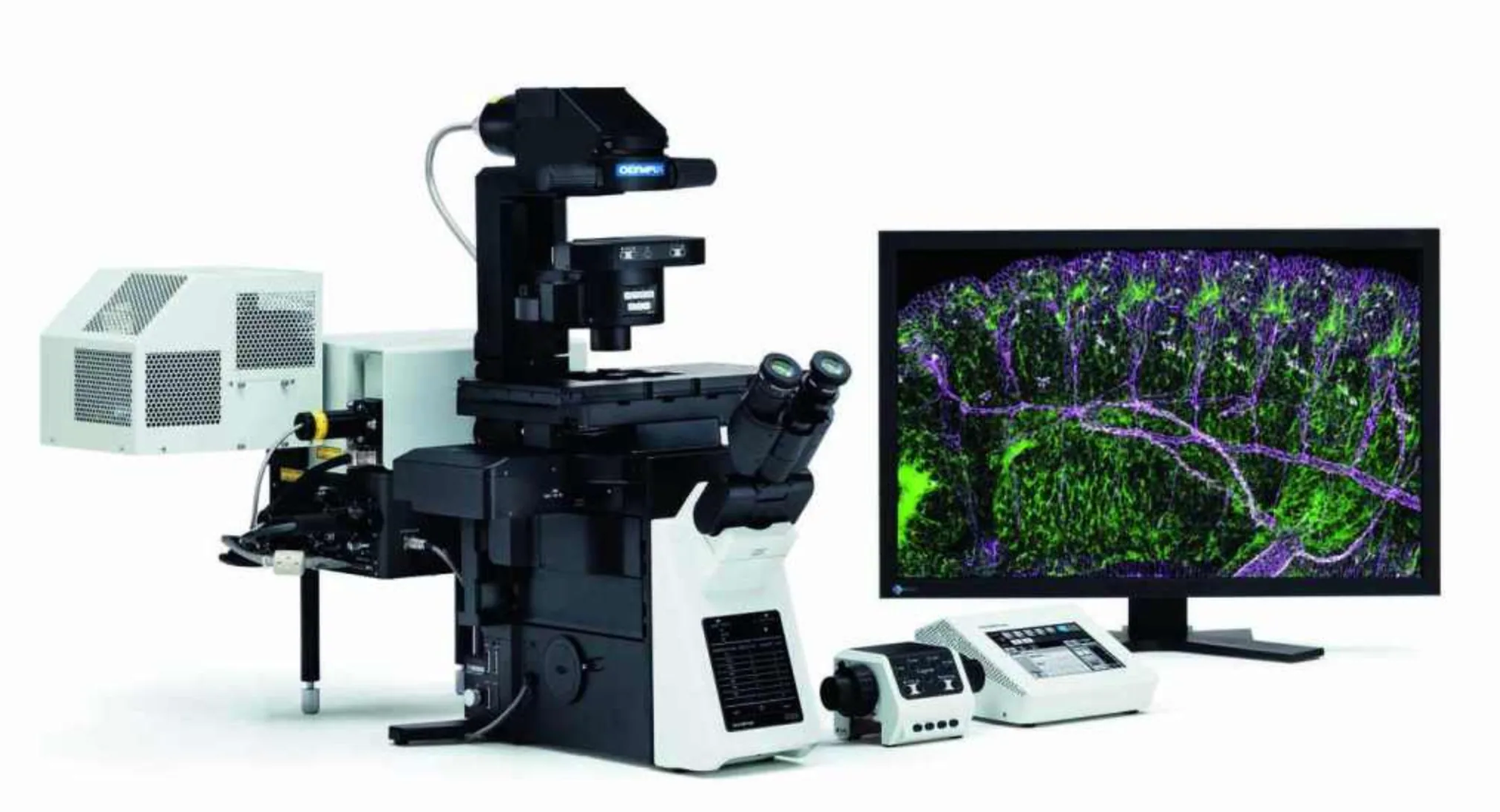
Confocal Microscopy (OLYMPUS FV1200)

Confocal Microscopy (OLYMPUS FV1200)

Confocal microscopy enables the acquisition of optically sectioned images with high resolution and minimal background noise. It is perfect for 3D tissue reconstruction, detailed colocalization studies, signal measurement in thick sections or individual cells, and live cell imaging. The system supports spectral detection from blue to infrared, features a motorized stage, phase contrast, and advanced capabilities, including FRAP, FRET, temperature and gas control, microfluidics, and electrical stimulation.

Full support is offered for image acquisition, quantitative analysis, and experimental design.

Users must complete theoretical and practical training and demonstrate proficiency before being granted autonomous access to the system.
Epifluorescence Microscopy (OLYMPUS IX71)

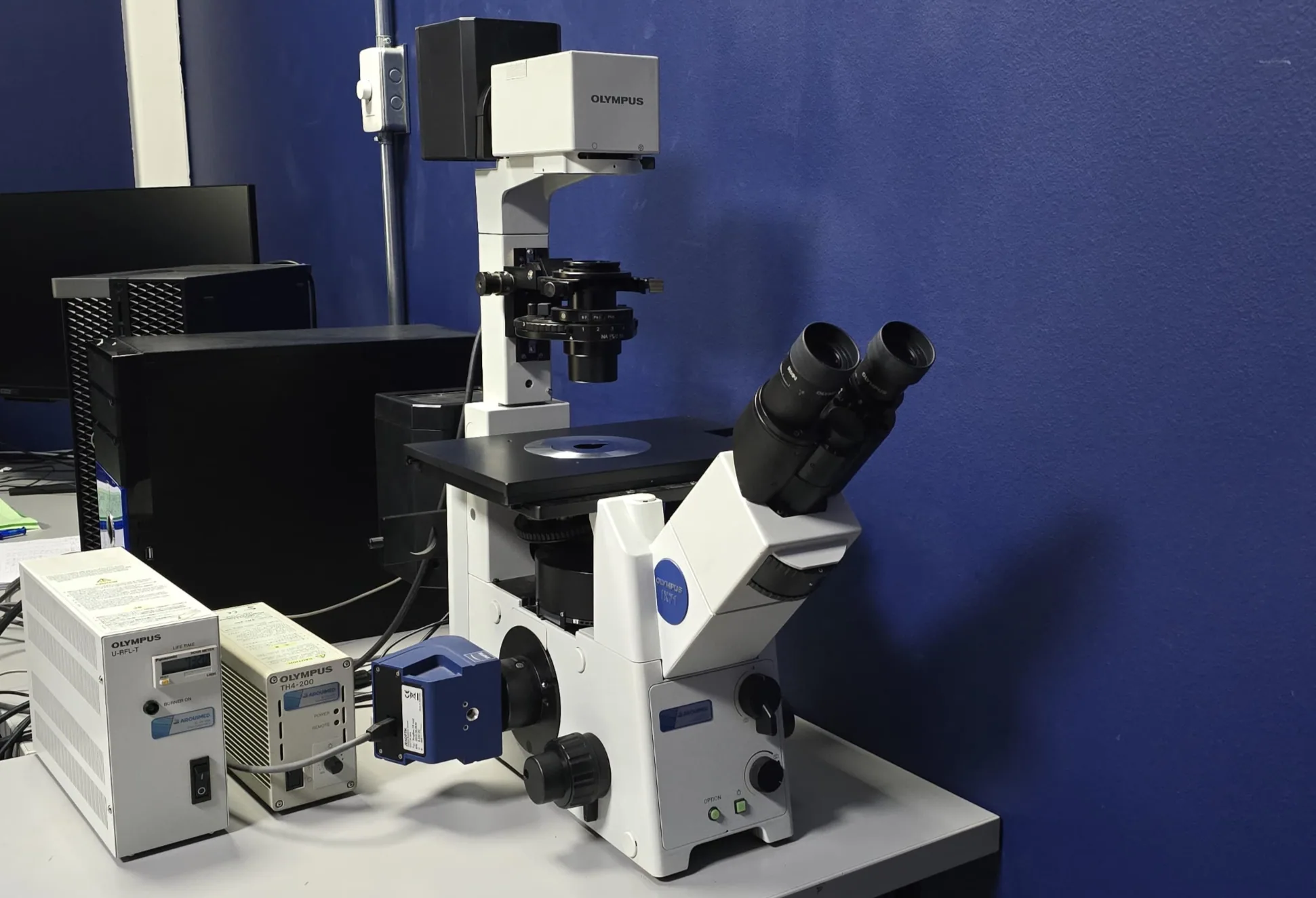
Epifluorescence Microscopy (OLYMPUS IX71)

This inverted optical system allows the visualization and documentation of biological processes in both fixed and live samples using fluorescent markers and/or conventional stains. It is commonly used for studies involving abundance, localization, colocalization, morphology, gene expression, and cell viability, among other applications. The system includes multiple excitation/emission channels, allowing the simultaneous detection of various fluorescent markers.

Users must complete theoretical and practical training and demonstrate proficiency before being granted autonomous access to the system.

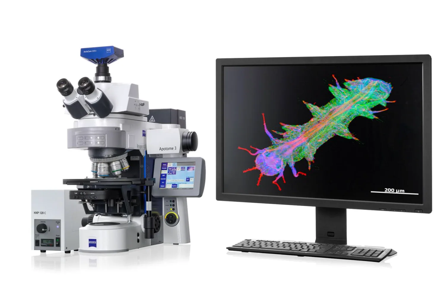
Stereological Microscopy (ZEISS Axio Imager M2)

Stereological Microscopy (ZEISS Axio Imager M2) This tool is designed for unbiased morphological quantification of three-dimensional structures from two-dimensional histological sections. It is widely used for accurate estimation of cell number, volume, surface area, and length in tissues, especially in neuroscience, histopathology, and structural morphometry. The system features a motorized stage. Support is provided for stereological sampling design and statistical data analysis. Users must complete theoretical and practical training and demonstrate proficiency before being granted autonomous access to the system.